Case of the Month ...

Case History
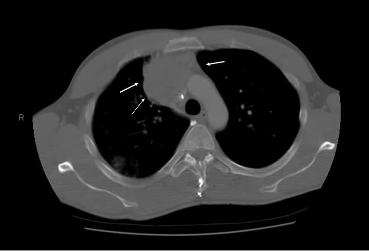
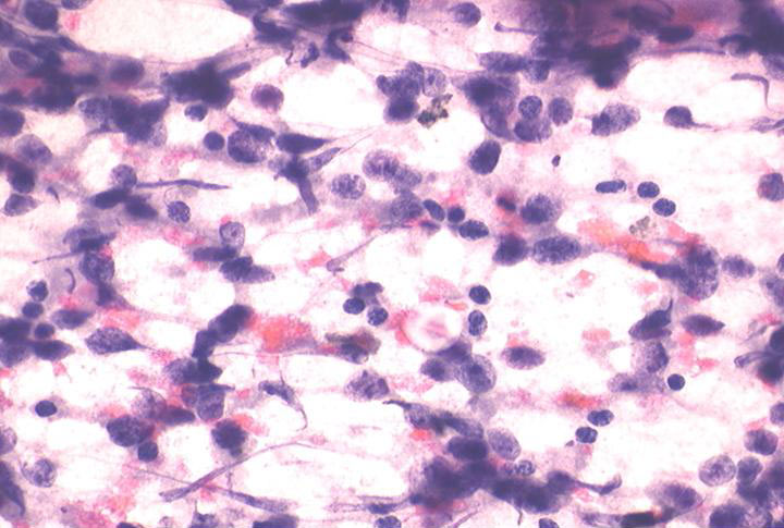
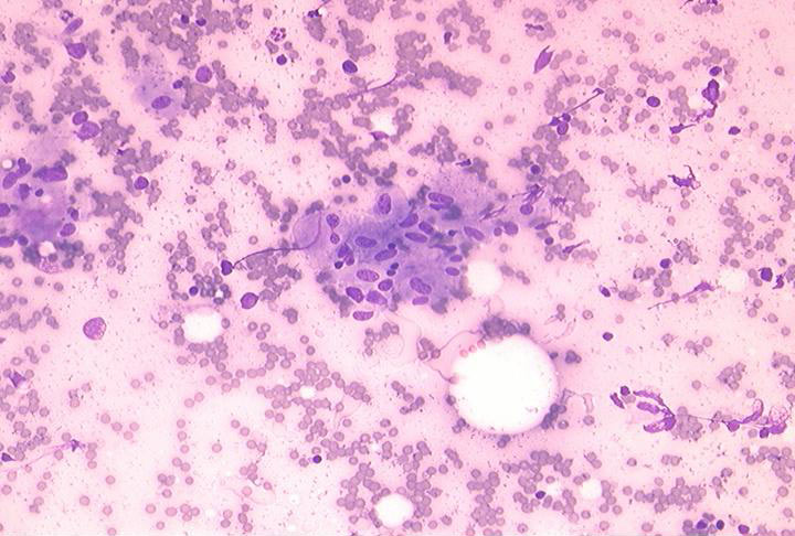
36-year-old male presented with a three-month history of back pain and ataxia. A chest radiograph demonstrated a right hilar mass. Chest CT demonstrated a mass measuring 5.3 x 5.2 cm in the right mediastinum. (Figure 1) A CT guided fine-needle aspiration was performed. The Papanicalaou stained slide revealed a cellular smear consisting of fragile large, round, discohesive cells with scant cytoplasm. (Figure 2) The nuclei had finely granular chromatin with variably sized nucleoli. (Figure 3) Histiocytes, giant cells, lymphocytes, plasma cells, and granulomas were present in the background. (Figure 4)
Check back in late November for the answer
Diagnosis & Discussion
click on image for larger version
CYTOLOGIC FINDINGS
The smears were hypercellular with loosely cohesive groups and single cells (Figure 1). The tumor cells had spindled or round to oval nuclei with marked pleomorphism, evenly distributed chromatin and abundant granular cytoplasm (Figure 2). Naked spindled nuclei were noted in the hemorrhagic background. The cell groups had the tendency to form acinar structures. A helpful feature was the presence of reddish cytopalasmic granules on Romanowsky stain (Figure 3, arrow). Intranuclear cytoplasmic inclusions were easily identified (Figure 4, arrow). Additional materials were sent for electron microscopy examination which showed numerous cytoplasmic membrane bound dense granules (Figure 5).
DIAGNOSIS: CAROTID BODY TUMOR - PARAGANGLIOMA
HISTOLOGIC FINDINGS
At excision, a lobulated brown 5 cm mass with areas of hemorrhage was noted. Well defined cell nests of ovoid to spindled shaped chief cells with finely granular chromatin and abundant eosinophilic cytoplasm were surrounded by sustentacular cells, and embedded in a well vascularized stroma (Figure 6). Immunohistochemical studies showed positivity for neuron-specific enolase (Figure 7A), chromogranin (7B) and synaptophysin (7C), in the cell nests and positivity for S-100 protein (7D) in the sustentacular cells.
DISCUSSION
Carotid body tumor is an uncommon lesion situated at the bifurcation of the common carotid artery with the usual presentation of a slow growing mass in the anterior triangle of the neck. A bruit can commonly be heard. Most of these tumors are benign with a 10% incidence of malignancy, evidenced by distant metastases.
Complications from fine needle aspiration have been reported including hemorrhage within the tumor in addition to one reported fatality from carotid thrombosis and cerebral embolism.
Differential diagnosis on fine needle aspiration includes primary neurogenic tumors and thyroid neoplasms. Aspiration findings in schwannomas show mainly cohesive groups of spindled to ovoid shaped cells, some wavy in appearance with sharply pointed ends. Nuclear palisading can be seen. The cells are often embedded in a collagenous matrix that appears fibrillary. S-100 protein immunostaining shows more diffuse positivity than in paragangliomas. Follicular carcinomas of the thyroid form similar acinar arrangements yet the nuclei are more round and uniform. Papillary adenocarcinomas of the thyroid gland commonly contain intranuclear cytoplasmic inclusions, however other cytologic features such papillary structures, nuclear grooves and nuclear overlapping are prominent. It is important to note that rare cases of primary thyroid paraganglioma have been reported.
The treatment or paragangliomas is usually embolization followed by surgical excision. Malignancy evidenced by metastases is noted in 6-10%, with 50% to lymph nodes and the remainder to lung and bone.
REFERENCES
1. Gonzalez-Campora R, Otal-Salaverri C, Panea-Flores P et al. Fine needle aspiration cytology of paraganglionic tumors. Acta Cytol 1988;32:386-390.
2. Hood IC, Qizilbash AH, Young JE et al. Fine needle aspiration biopsy cytology of paragangliomas. Cytologic, light microscopic and ultrastructural studies of three cases. Acta Cytol 1983;27:651-657.
3. Engzell V, Franzen S, Zajicek J. Aspiration biopsy of tumors of the neck:II. Cytologic findings in 13 cases of carotid body tumors. Acta Cytol 1971; 15:25 -30.
4. Vodovnik A. Fine needle aspiration cytology of primary thyroid paraganglioma. Acta Cytol 2002;46:1133-1137.
5. Zaharopoulos P. Diagnostic challenges in the fine-needle aspiration diagnosis of carotid body paragangliomas: report of two cases. Diagn Cytopathol 2000;23:202-207.
6. Capella C, Riva C, Cornaggia M, Chiaravalli AM, Frigerio B. Histopathology, Cytology and Cytochemistry of pheochromocytomas and paragangliomas including chemodectomas. Path Res Pract 1988;183:176-187.
ACKNOWLEDGEMENT
This case is contributed by Dr. Joan Cangiarella from New York University, New York, NY.