Case of the Month ...
Case History
A 51 year old female was incidentally found to have a large right renal mass on CT scan while hospitalized for COPD related clinical symptoms. A CT -guided Fine needle aspiration of the right renal mass was requested to rule out renal cell carcinoma.
Diagnosis & Discussion
click on image for larger version
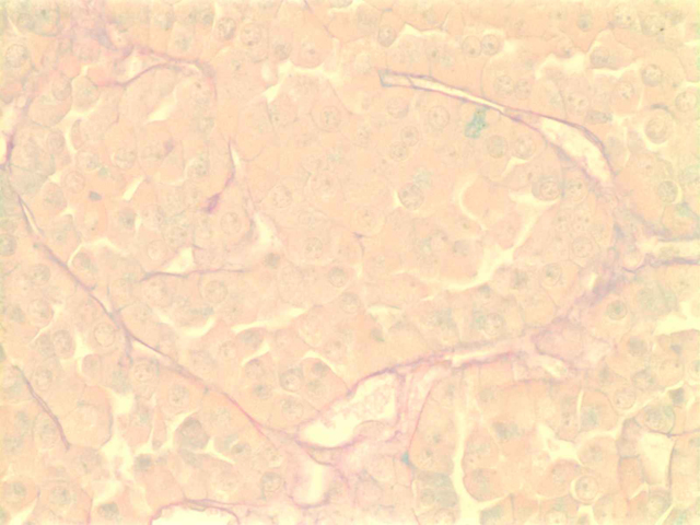
Figure 1
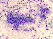
Image figures:
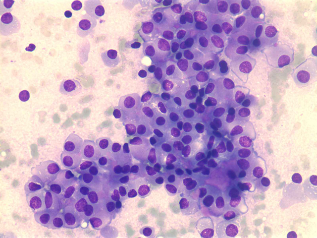
- Figure 1: FNA cytology smear, Diff quick stain, X 100
- Figure 2: FNA cytology smear, Diff quick stain, X 100
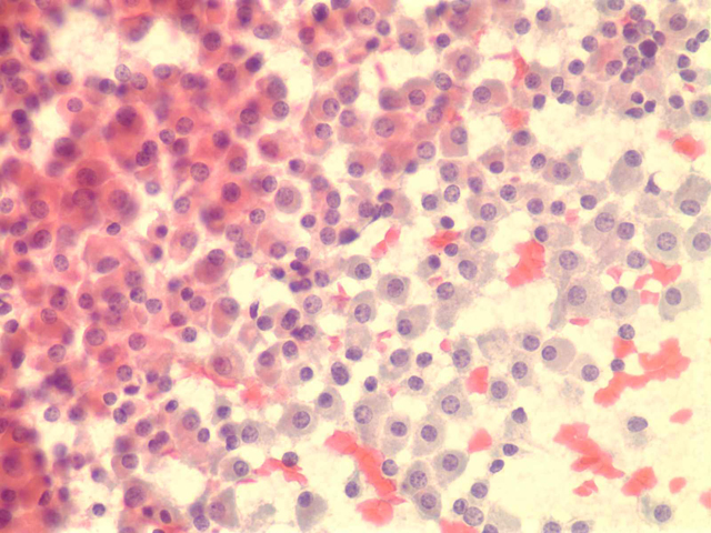
- Figure 3: FNA cytology smear, Diff quick stain, X 400
- Figure 4: FNA cytology smear, Papanicolaou stain, X 100
- Figure 5: FNA cytology smear, Papanicolaou stain, X 100
- Figure 6: FNA cytology smear, Papanicolaou stain, X 100
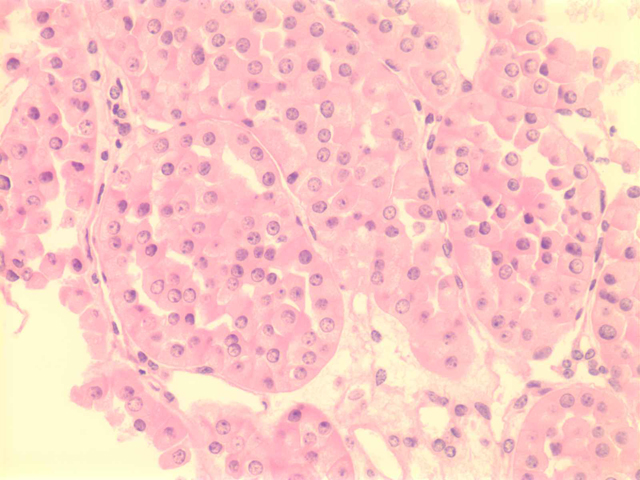
- Figure 7: FNA cell block, H & E stain, X 400
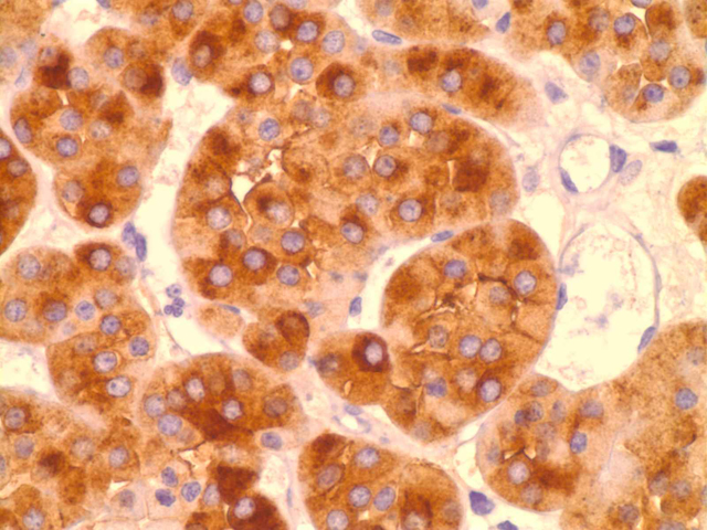
- Figure 8: FNA cell block, EMA immunoperoxidase stain, X 600
- Figure 9: FNA cell block, Colloidal iron stain, X 600
Diagnosis: Renal Oncocytoma.
Cytologic Findings:
- Very cellular aspirate with cohesive fragments and single cells.
- Cells are monotonous and round to polygonal
- Abundant granular cytoplasm with occasional small vacuoles.
- Nuclei are small and monotonous with finely granular nuclear chromatin.
- Nucleoli are inconspicuous
- No cellular atypia, mitotic figures or necrosis are identified.
Discussion:
Renal oncocytoma is a benign renal neoplasm which comprises 3-7 percent of adult kidney tumors. One half to two-thirds of these lesions are found incidentally. However, they can present with hematuria, flank pain and be palpable on clinical examination. Occasionally large bilateral tumors can present as renal failure. Oncocytomas have been reported in patients with Birt-Hogg-Dube and Von-Hippel Lindau syndromes. The most common cytogenetic abnormalities include loss of chromosomes 1 and y and translocations of 11q13. In very rare cases, both kidneys show diffuse involvement with macroscopic and microscopic oncocytoma, chromophobe renal cell carcinomas, and hybrid tumors with oncocytic changes of the adjacent renal tubules epithelium. These cases have been termed renal oncocytosis.
The major differential diagnosis to consider is chromophobe renal cell carcinoma. Compared to oncocytoma, chromophobe renal cell carcinoma shows nuclear hyperchromasia with nuclear membrane irregularities. There are prominent perinuclear halos, and greater nuclear variability. Colloidal iron will be diffusely positive in chromophobe renal cell carcinoma while EMA is negative, in contrast to oncocytoma where colloid iron is negative and EMA is positive.
References:
- Gudjartsson T, Sverrir H, et al: Renal oncocytoma: a clinicopathological analysis of 45 consecutive cases. British Journal of Urology, 2005, 96, 1275-1279.
- Campodonico F, Carmignani G, Tocini C.: Bilateral renal oncocytosis with renal failure. Archives of Pathology and Laboratory medicine. 2001, 125(5). 648-649.
- Christian P, Walther M, et al: Renal tumors in the Birt-Hogg-Dube syndrome. American Journal of Surgical Pathology. 2002, 26(12), 1542-1552.
- Fiske J, Patel R, et al.: Multifocal renal oncocytoma in a patient with Von Hippel-Lindau mutation. 2005, 66(6), 1320.
- Gladell P, Lindgren V, et al.: High incidence of chromosome 1 abnormalities in a series of 27 renal oncocytomas, Archives of Pathology and Laboratory medicine. 2007, 131, 81-85
- Tickoo S, Victor E, Mahul A, Srigley J, Epstein J: Renal Oncocytosis: a morphologic study of fourteen cases. The American Journal of Surgical Pathology. 1999, 23(9), 1094.
Acknowledgement: This case is submitted by Jeremiah Watkins M.D. and Momin T. Siddiqui M.D., FIAC. Department of Pathology and Laboratory Medicine, Emory University Hospital , Atlanta , GA.