Case of the Month ...
Clinical History:
73 year old male status post treatment for adenocarcinoma of the lung is found to have an 8cm well defined mass in the liver. Patient has no known cirrhosis, but has long history of alcohol abuse and undergoes CT-guided FNA of the mass.
Diagnosis & Discussion
click on image for larger version
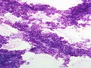
Figure 4
Figures 1-4:
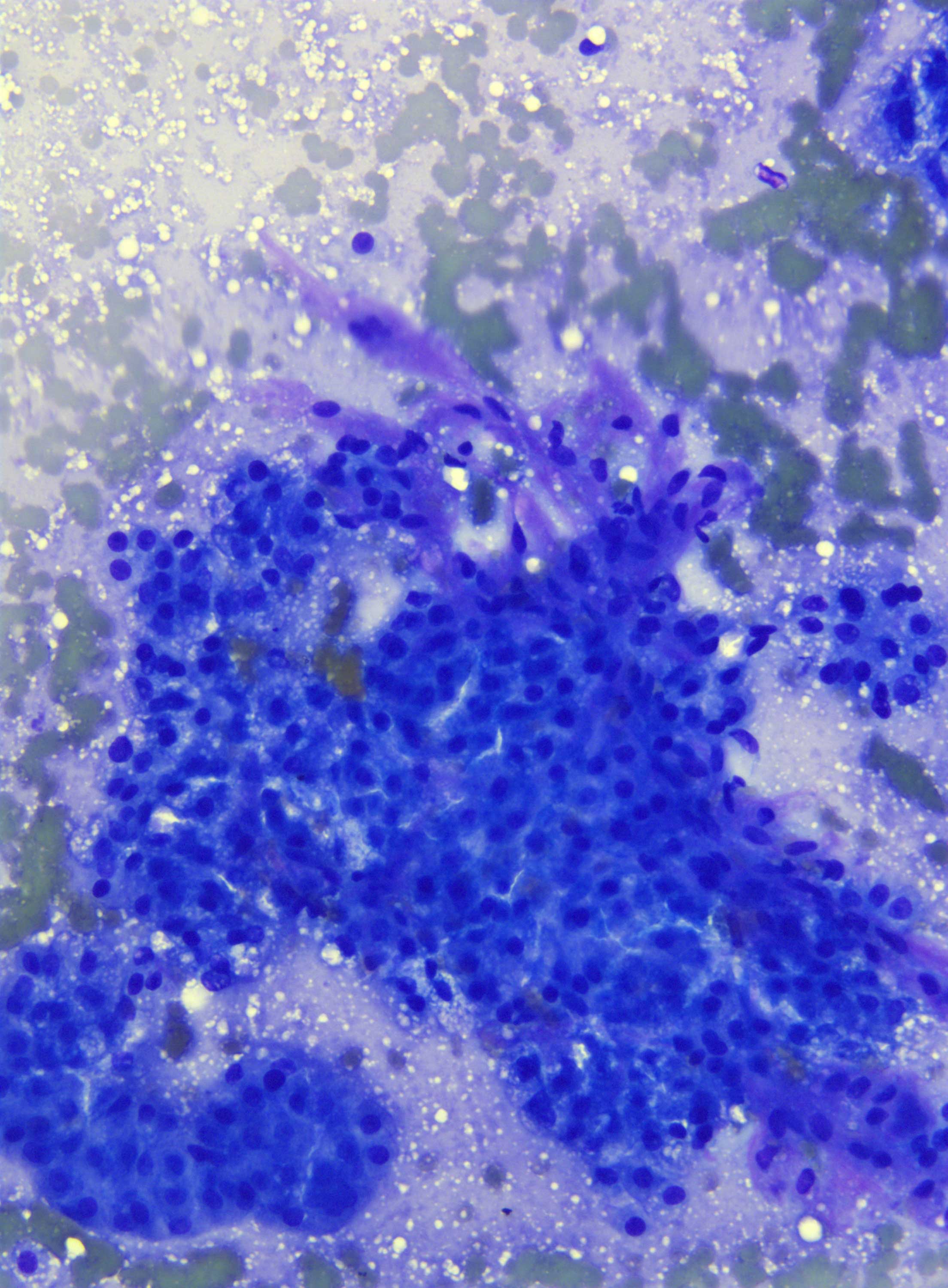
Figure 1: Fine needle aspiration, liver mass, Diff Quik stain, low magnification
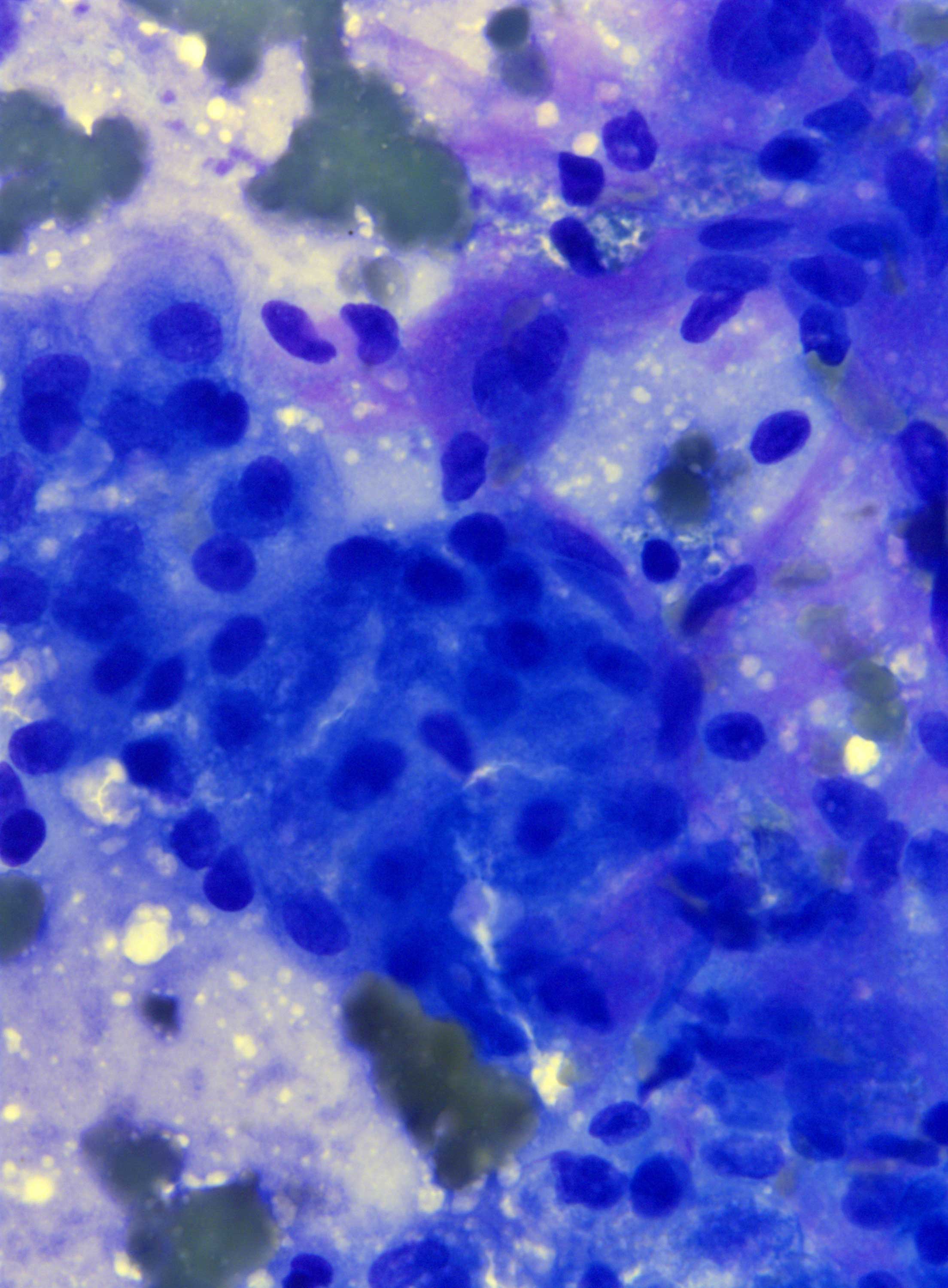
Figure 2: Fine needle aspiration, liver mass, Diff Quik stain, high magnification
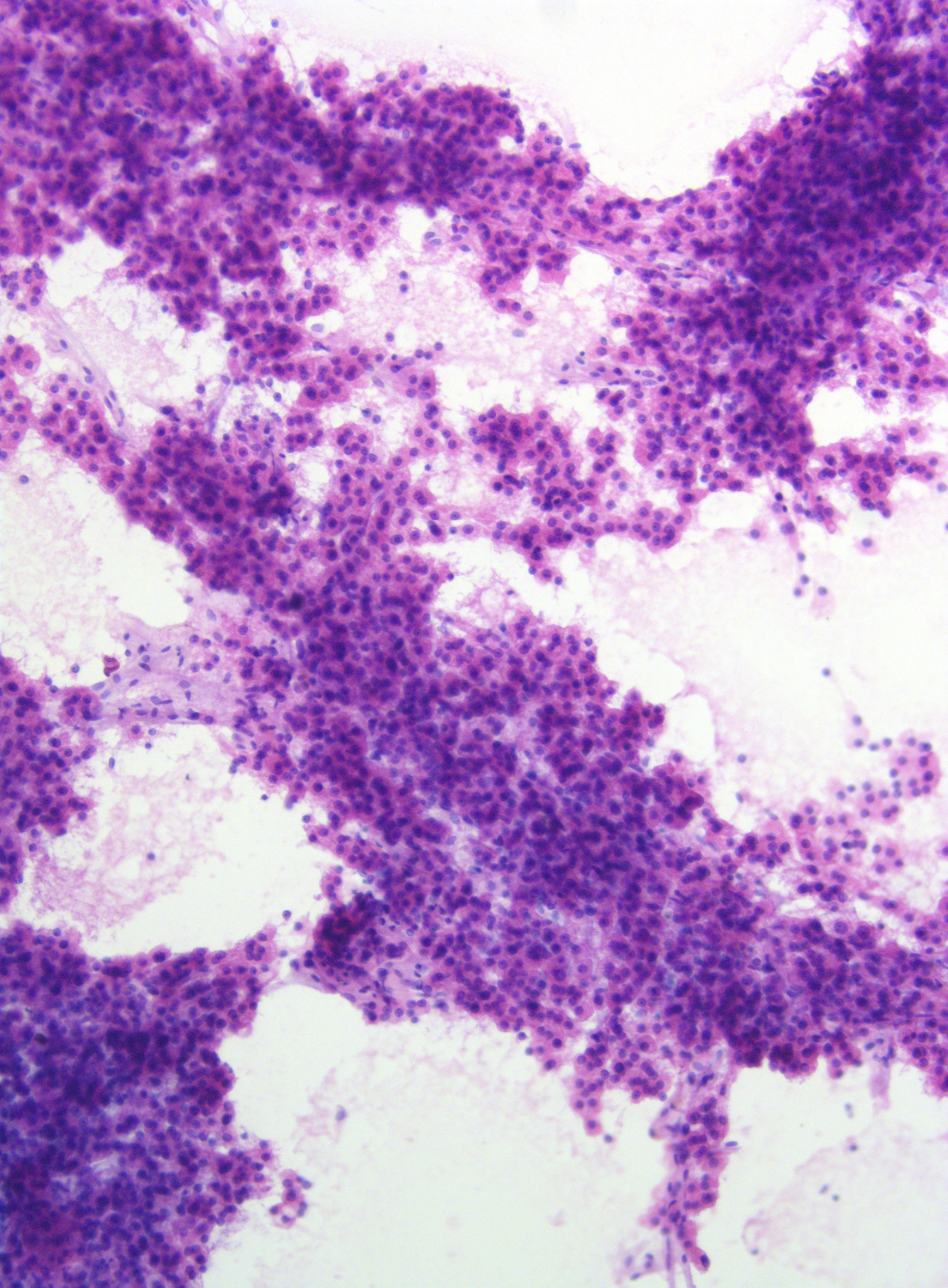
Figure 3: Fine needle aspiration, liver mass, H&E stain, low magnification
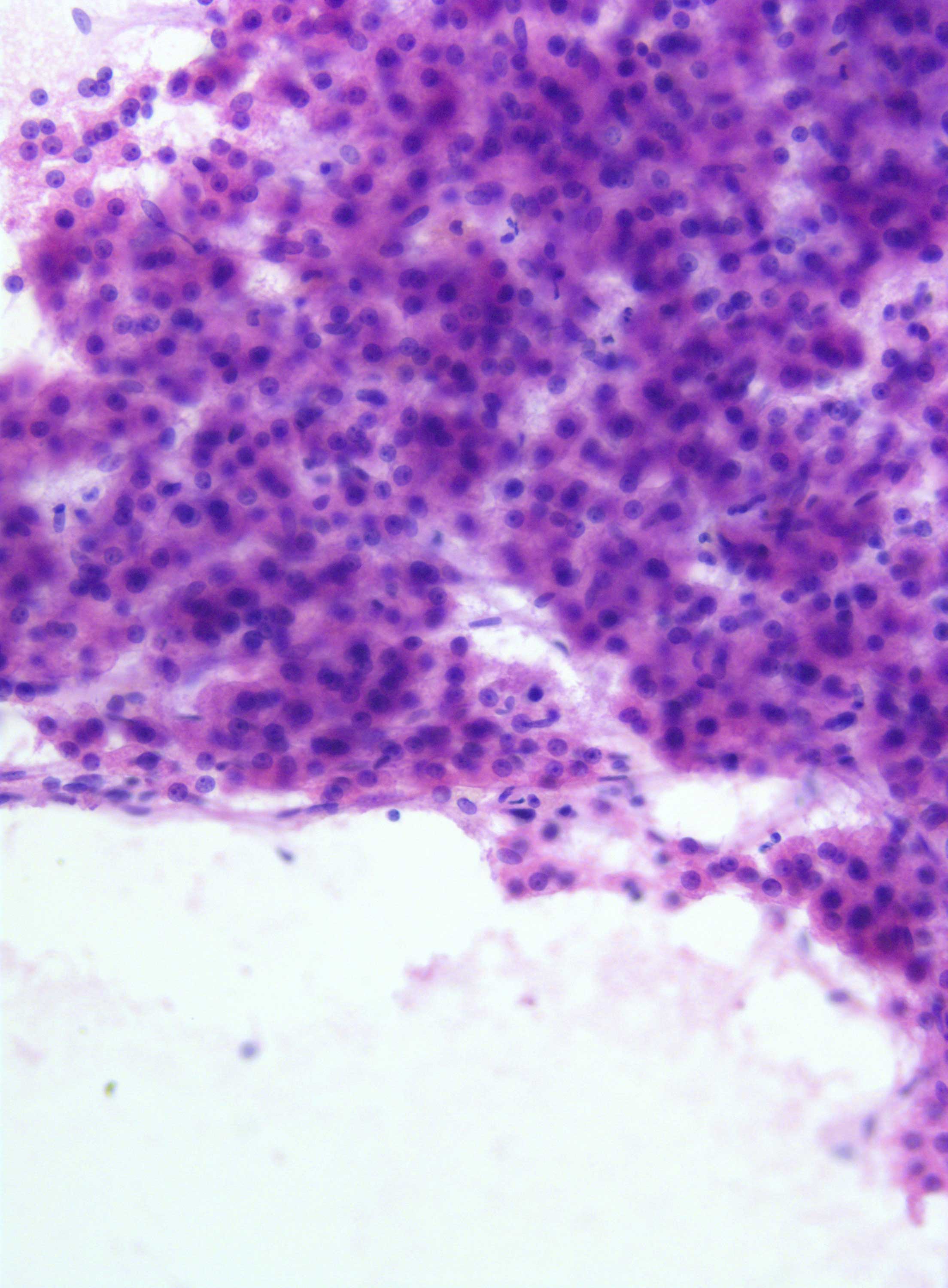
Figure 4: Fine needle aspiration, liver mass, H&E, high magnificationQuestions:
- What is the most likely diagnosis?
- Non-lesional liver
- Regenerative Nodule
- Well differentiated hepatocellular carcinoma
- Metastatic carcinoma
- Cholangiocarcinoma
- Which feature is most suggestive of hepatocellular carcinoma?
- Anisonucleosis
- Transgressing vessels
- Granular cytoplasm
- Nucleoli
- Pleomorphism
- Which immunohistochemical stain would be the most useful in this case?
- MOC-31
- Hep Par 1
- CEA
- TTF-1
- Glypican -3
Discussion:
Cytology preparations show a cellular aspirate comprised of flat groups of monotonous, somewhat bland appearing cells with central nuclei and eosinophilic cytoplasm in a clean background. The cells are hepatoid with diffuse minimal increase in the nuclear to cytoplasmic ratio. Transgressing vessels are easily identified. Ductal cells are not appreciated. These findings are compatible with a well differentiated hepatocellular carcinoma.
Hepatocellular carcinoma is the sixth most common type of cancer and the third most common cause of death [1]. Typically it is found in a background of cirrhosis and distinction of well differentiated carcinoma from the background liver can be challenging [2-4]. Cytologic features of hepatocellular carcinoma include monomorphic , small hepatocytes with increase in nuclear to cytoplasmic ratio, endothelial cell wrapping, transgressing vessels, and lack of ductal cells[5-8]. Of these the endothelial cell wrapping and transgressing vessels have a sensitivity of 94% and a specificity of 91% [7]. Positivity for glypican-3 is helpful in distinguishing well differentiated hepatocellular carcinomas from benign hepatic nodules [9-11] as is loss of the reticulin framework [12]. In cases of more poorly differentiated carcinomas, Hep Par 1 positivity is helpful to distinguish lineage; Hep Par 1 will not distinguish benign from malignant hepatocytes [13]. Hepatocellular carcinomas are highly heterogeneous tumors with regard to degree of differentiation, histologic pattern, and cell morphology. Image guided fine needle aspiration cytology overall has a high specificity and sensitivity with low morbidity [14-16].
Answers:
1: C
2: B
3: EReferences:
- Ferlay, J., et al., Estimates of worldwide burden of cancer in 2008: GLOBOCAN 2008. Int J Cancer, 2010. 127(12): p. 2893-917.
- Schlageter, M., et al., Histopathology of hepatocellular carcinoma. World J Gastroenterol, 2014. 20(43): p. 15955-64.
- Hytiroglou, P., et al., Hepatic precancerous lesions and small hepatocellular carcinoma. Gastroenterol Clin North Am, 2007. 36(4): p. 867-87, vii.
- Roncalli, M., Y.N. Park, and L. Di Tommaso, Histopathological classification of hepatocellular carcinoma. Dig Liver Dis, 2010. 42 Suppl 3: p. S228-34.
- Kung, I.T., S.K. Chan, and K.H. Fung, Fine-needle aspiration in hepatocellular carcinoma. Combined cytologic and histologic approach. Cancer, 1991. 67(3): p. 673-80.
- Lin, C.C., et al., Fine-needle aspiration cytology to distinguish dysplasia from hepatocellular carcinoma with different grades. J Gastroenterol Hepatol, 2008. 23(7 Pt 2): p. e146-52.
- Pitman, M.B. and W.M. Szyfelbein, Significance of endothelium in the fine-needle aspiration biopsy diagnosis of hepatocellular carcinoma. Diagn Cytopathol, 1995. 12(3): p. 208-14.
- Sole, M., et al., Value and limitations of cytologic criteria for the diagnosis of hepatocellular carcinoma by fine needle aspiration biopsy. Acta Cytol, 1993. 37(3): p. 309-16.
- Zhu, Z.W., et al., Enhanced glypican-3 expression differentiates the majority of hepatocellular carcinomas from benign hepatic disorders. Gut, 2001. 48(4): p. 558-64.
- Nakatsura, T., et al., Glypican-3, overexpressed specifically in human hepatocellular carcinoma, is a novel tumor marker. Biochem Biophys Res Commun, 2003. 306(1): p. 16-25.
- Capurro, M., et al., Glypican-3: a novel serum and histochemical marker for hepatocellular carcinoma. Gastroenterology, 2003. 125(1): p. 89-97.
- Bergman, S., F. Graeme-Cook, and M.B. Pitman, The usefulness of the reticulin stain in the differential diagnosis of liver nodules on fine-needle aspiration biopsy cell block preparations. Mod Pathol, 1997. 10(12): p. 1258-64.
- Lamps, L.W. and A.L. Folpe, The diagnostic value of hepatocyte paraffin antibody 1 in differentiating hepatocellular neoplasms from nonhepatic tumors: a review. Adv Anat Pathol, 2003. 10(1): p. 39-43.
- Kuo, F.Y., et al., Fine needle aspiration cytodiagnosis of liver tumors. Acta Cytol, 2004. 48(2): p. 142-8.
- Longchampt, E., C. Patriarche, and M. Fabre, Accuracy of cytology vs. microbiopsy for the diagnosis of well-differentiated hepatocellular carcinoma and macroregenerative nodule. Definition of standardized criteria from a study of 100 cases. Acta Cytol, 2000. 44(4): p. 515-23.
- Pupulim, L.F., et al., Algorithm for immediate cytologic diagnosis of hepatic tumors. AJR Am J Roentgenol, 2008. 190(3): p. W208-12.
Contributed by:
Dianne Grunes, MD
Assistant Professor of Pathology
Department of Pathology
University of Mississippi Medical Center, Jackson, MS