Case of the Month ...
An 85-year-old man with a history of coronary artery disease, hypertension, hyperlipidemia, and asthma, presented with painless jaundice and recent unintentional weight loss. Blood work up was notable for abnormal hepatic function test, with elevated total bilirubin 12.1 mg/dl (normal 0.0-1.3mg/dL). CT abdomen imaging showed a 2.1 cm mass in the distal common bile duct causing severe upstream biliary dilation. Gross examination of the voided urine collected was notable for amber-brown color.
Authors
- Simon Sung, MD, Assistant Professor, Columbia University Irving Medical Center, Dept of Cell Biology and Pathology, New York NY
Diagnosis & Discussion
click on image for larger version
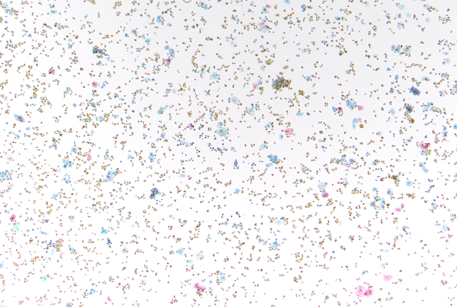
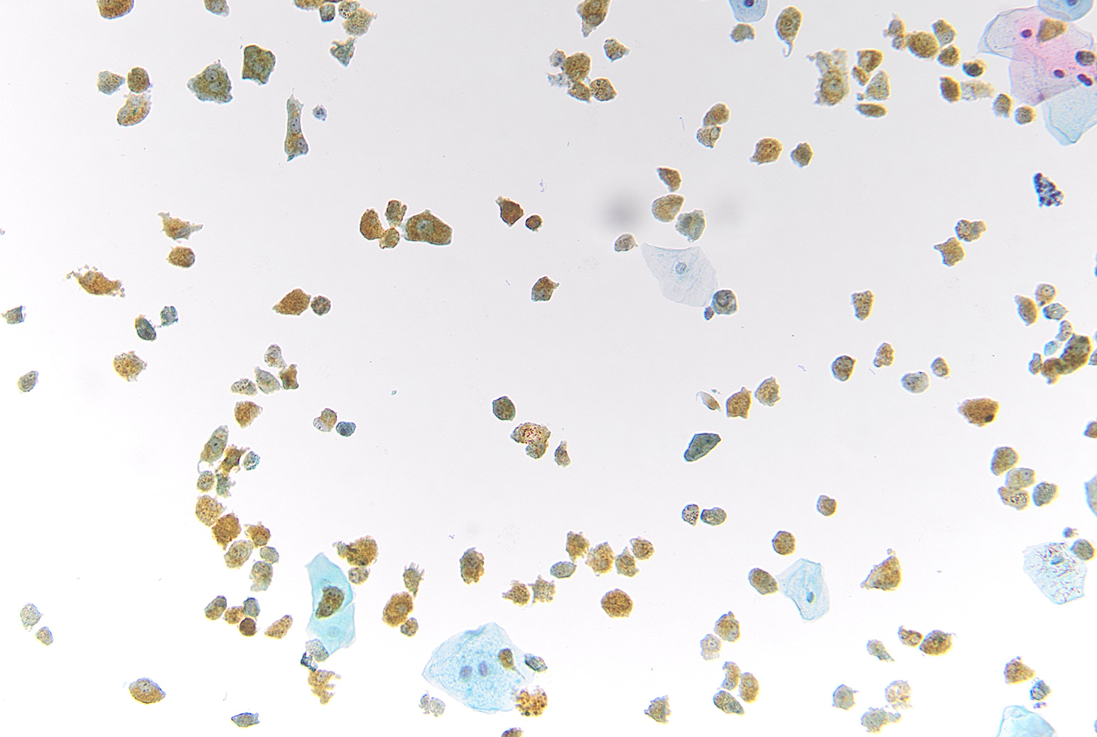
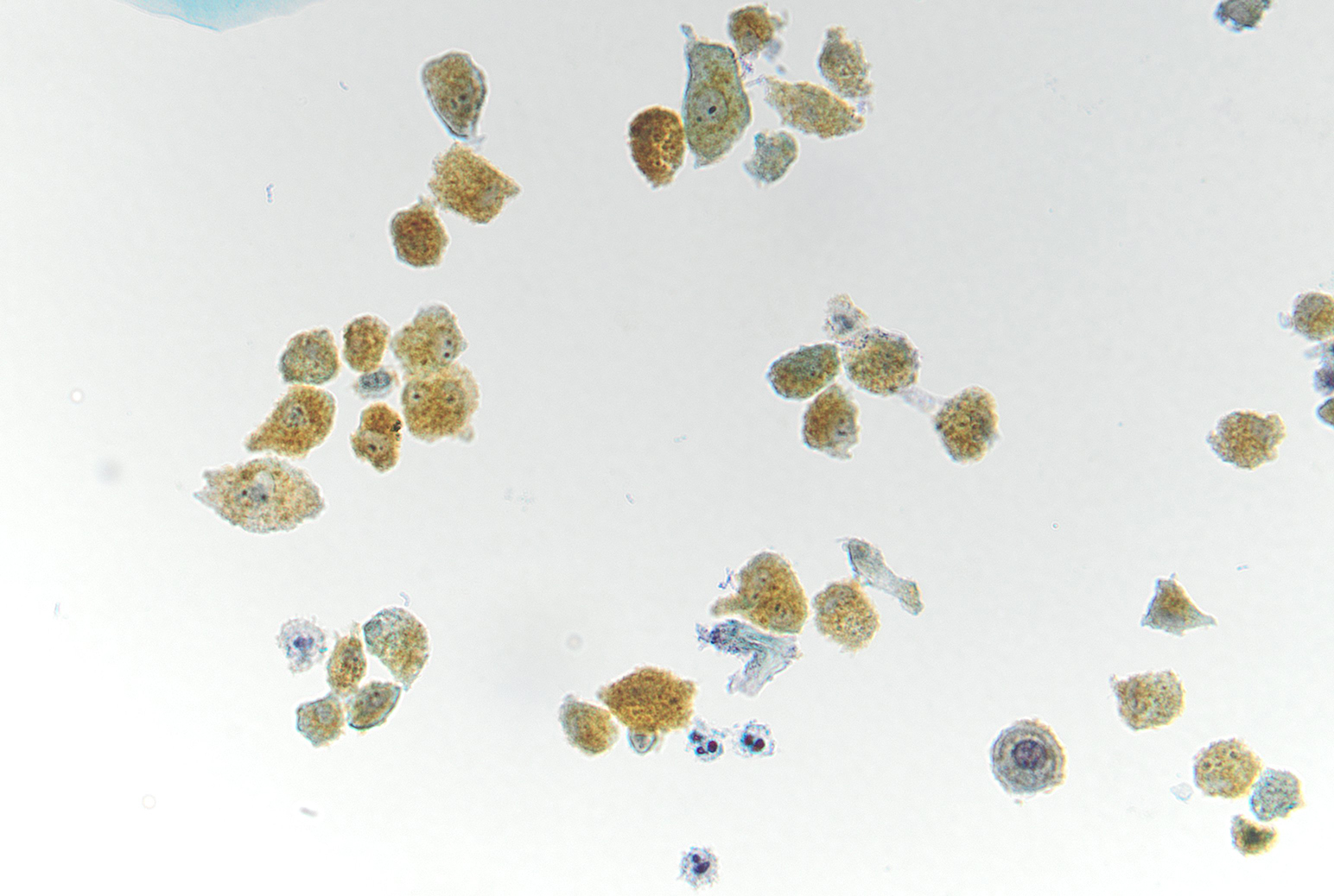
Figure 3 Images 1-3:
- Figure 1: ThinPrep 40x
- Figure 2: ThinPrep 200x
- Figure 3: ThinPrep 400x
Questions:
- What pigment is in the cytoplasm?
- Hemosiderin
- Melanin
- Lipofuscin
- Bilirubin
- Bile
- Based on patient’s presentation and cytomorphologic findings, what is the most likely cause of the pigmented cells in the urine?
- Undiagnosed melanoma
- Nephrolithiasis
- Severe biliary obstruction, leading to increased conjugated bilirubin in the blood which is excreted through the kidney
- Bladder outlet obstruction
Answers:
Question 1: Correct answer is D
The low and high magnifications of the figures show yellow gold, coarse pigment, which is most consistent with bilirubin. Typically, on Papanicolaou stain, melanin is dusty fine brown, hemosiderin is chunky dark brown, lipofuscin is gold and normally related to cellular aging, and bile is dark green, including on Romanowsky stain. The patient’s presentation of painless jaundice and hyperbilirubinemia is most compatible with presence of bilirubin in the urine, leading to the gross appearance of amber/brown (“tea colored”) urine and presence of extensive pigmented cells.Question 2: Correct answer is C
The presence of bilirubin in urine is unusual. This patient was found to have a large obstructing mass in the distal common bile duct and ultimately diagnosed with cholangiocarcinoma. Dark tea-colored urine indicates presence of conjugated bilirubin (water soluble bilirubin, that is excreted through the kidney) and would usually indicate liver or biliary disease.References:
- Hoilat GJ, John S. Bilirubinuria. 2023 Aug 8. In: StatPearls [Internet]. Treasure Island (FL): StatPearls Publishing; 2023 Jan–. PMID: 32491371.
- Cibas, ES, Ducatman, BS. Cytology: diagnostic principles and clinical correlates. Philadelphia, PA: Elsevier; 2019.
- O'Connell W, Shah J, Mitchell J, Prologo JD, Martin L, Miller MJ Jr, Martin JG. Obstruction of the Biliary and Urinary System. Tech Vasc Interv Radiol. 2017 Dec;20(4):288-293. doi: 10.1053/j.tvir.2017.10.010. Epub 2017 Oct 9. PMID: 29224663.
- Patel T. Cholangiocarcinoma--controversies and challenges. Nat Rev Gastroenterol Hepatol. 2011 Apr;8(4):189-200. doi: 10.1038/nrgastro.2011.20. PMID: 21460876; PMCID: PMC3888819.