Case of the Month ...

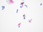
A 41-year-old male with a history of Crohn’s disease (in remission) and on HIV preexposure prophylaxis, presents to his primary physician’s office with complaints of mild discomfort during defecation. Laboratory tests for trichomonas, gonorrhea, syphilis and HIV were negative. A liquid-based anal Pap test was performed and a ThinPrep slide was prepared and Papanicolaou stained. Figures 1-3 show what was observed by the attending cytopathologist. The objects of interest ranged in size from 10-12 micrometers. A stool ova and parasite examination from the patient was not submitted.
Authors
Lagnajita Datta, MD
University Hospitals Cleveland Medical Center and Case Western Reserve University, Cleveland, Ohio
click on image for larger version
Figure 3 Images 1-3:
Figure 1: Papanicolaou-stained ThinPrep at 400X magnification
Figure 2: Papanicolaou-stained ThinPrep at 600X magnification
Figure 3: Papanicolaou-stained ThinPrep at 600X magnificationQuestions:
- What is the most likely diagnosis?
- Cyst forms of Entamoeba histolytica
- Giardia intestinalis
- Cyst forms of Iodamoeba buetschlii
- Trichomonas spp.
- What special stain can be used to confirm the diagnosis?
- Perl’s Prussian blue (iron) stain
- Iodine stain
- Oil Red O stain
- Silver stain
Answers:
Question 1: Correct answer is D. Cyst forms of Iodamoeba buetschlii.
I. butschlii is a nonpathogenic amoeba found worldwide and is a commensal parasite of the large intestine in humans. They are generally recovered from stool ova and parasite examinations when cysts and trophozoites of I. butschlii are shed. The mode of transmission is by ingestion of infective cysts in contaminated food or water.
A characteristic morphologic feature of this organism is its compact, large, discrete glycogen mass, and single nucleus with a large central karyosome. Cysts measure 5–20 microns (usual range 10–12 microns) and may be oval or circular. On the Papanicolaou-stained ThinPrep slide, the glycogen mass did not stain but appeared as a relatively clear well-defined area within the cytoplasm. There appears to be no definitive association between I. butschlii and HIV infection. However, men who have sex with men (MSM) are more likely to have I. butschlii in the stool than the non-MSM male population.
It is imperative to distinguish I. butschlii from E. histolytica due to the pathogenicity of the latter. Organism size may overlap between I. butschlii and E. histolytica; however, the E. histolytica nucleus contains a smaller karyosome and peripheral rim of condensed chromatin. Although cytoplasmic inclusions may be seen in both organisms, I. butschlii does not contain ingested red blood cells, as can be seen with E. histolytica.
Iodamoeba may also overlap in size with Giardia intestinalis, which has also been reported on anal Pap smears, but Iodamoeba lacks multiple nuclei, median bodies, and central axoneme seen with Giardia.
Iodamoeba differ from trichomonads in their circular to oval shape, as opposed to the piriform shape and the presence of flagella in trichomonads.
Question 2: Correct answer is B Iodine stain.
The name Iodamoeba is derived from the iodophilic glycogen mass which stains reddish brown on iodine preparation. The mass is typically referred to as a vacuole, but on electron microscopy studies, it is not membrane bound and therefore does not qualify as a true vacuole. An iodine preparation on an unstained PreservCyt-fixed ThinPrep slide can be performed if I. butschlii cysts are suspected and can serve as a confirmatory test.
References
- Garcia LS. Diagnostic medical parasitology. 5th ed. Washington, D.C.: ASM Press, American Society for Microbiology; 2007. p 27.
- Stark D, Fotedar R, Van Hal S, et al. Prevalence of enteric protozoa in human immunodeficiency virus (HIV)-positive and HIV-negative men who have sex with men from Sydney, Australia. Am J Trop Med Hyg 2007;76:549–552.
- Stensvold CR, Lebbad M, Clark CG. Last of the human protists: The phylogeny and genetic diversity of Iodamoeba. Mol Biol Evol 2012;29:39–42.
- Arava S, Sharma A, Iyer VK, Mathur SR. Iodamoeba butschlii in a routine cervical smear. Cytopathol 2010;21:342–343.
- Schuetz AN, Pritt BS, Schreiner AM. Iodamoeba butschlii in an anal pap test confirmed by iodine stain. Diagn Cytopathol. 2014 Sep;42(9):775-7. doi: 10.1002/dc.23042. Epub 2013 Oct 25. PMID: 24167099.

