Case of the Month ...
A 68-year-old female non-smoker with a medical history of type 2 diabetes mellitus, hyperlipidemia, and anxiety presented with multiple pulmonary nodules discovered on imaging. Radiology revealed a 20 x 20 x 20 mm right upper lobe nodule and a 12 x 8 mm right lower lobe nodule, which has been stable since 2023. Diagnostic evaluation via robotic navigation-guided transbronchial fine-needle aspiration (FNA) provided cytology material for smears, ThinPrep preparation, and a cell block. The patient’s history is significant for severe pneumonia eight years ago during a “Valley fever” outbreak in Tucson, Arizona.
Authors
- Amanda Felty, BS, Western University of Health Sciences, College of Osteopathic Medicine of the Pacific Northwest
- Qiuying Shi, MD. MS, Oregon University and Science University
click on image for larger version
Figure 3 Figure 4 Figure 5
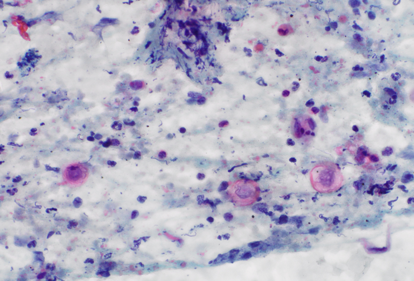
- Figure 1: Transbronchial FNA of right upper lobe lung nodule, Diff Quik smears, x400 magnification
- Figure 2: Transbronchial FNA of right upper lobe lung nodule, Papanicolaou smears, x400 magnification
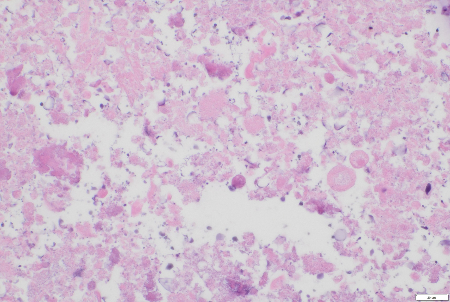
- Figure 3: Transbronchial FNA of right upper lobe lung nodule, cell block, H&E stain, x400 magnification
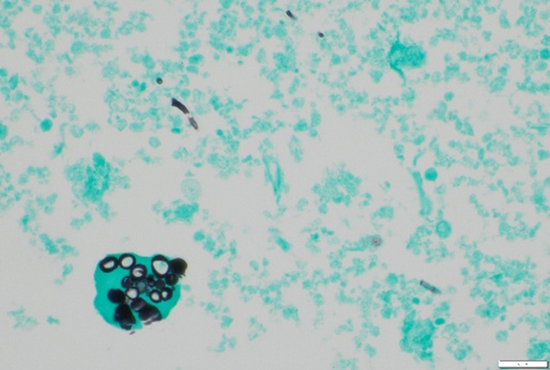
- Figure 4: Transbronchial FNA of right upper lobe lung nodule, cell block, GMS stain, x400 magnification
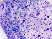
- Figure 5: Transbronchial FNA of right upper lobe lung nodule, cell block, PAS stain, x400 magnification
Questions:
- What is the most appropriate adequacy interpretation for this specimen?
- Non-diagnostic
- Benign; positive for organisms
- Atypical cell present
- Suspicious for malignancy
- Positive for malignancy
- What special stains are most useful for identifying the microorganisms shown in these cytologic specimens?
- GMS and PAS stains
- Mucicarmine
- AFB
- Calcofluor white only
- Potassium hydroxide preparation
- Which morphologic features are most characteristic of the spherules in the cytology specimen?
- Small 2-4 μm yeast forms within macrophages
- Large 20-200 μm spherules with thick walls containing endospores
- Septate hyphae with dichotomous branching
- Encapsulated yeast forms
- Broad-based budding yeast
Answers:
Question 1: Correct answer is B. Negative for malignancy; positive for organisms.
Smears and special stains demonstrate characteristic large spherules ranging from 20 to 200 μm with thick, doubly refractile walls containing internal endospores. The cytologic features include necrosis with microorganism-like structures and dense keratin-like material, which are typical findings in coccidioidomycosis. GMS and PAS stains highlight multiple fungal forms, including spherules with endospores, smaller yeast forms, and occasional hyphae. The identification of spherules in clinical specimens is considered diagnostic for coccidioidomycosis, even without positive culture results. The background shows necrotic debris with a paucity of inflammation, which is characteristic of pulmonary coccidioidomycosis in fine-needle aspiration specimens. Under the WHO Reporting System for Lung Cytopathology, infectious processes are classified within the "benign" category, with the diagnosis rendered as specifically as possible.
Option A (non-diagnostic) is incorrect because the specimen contains identifiable diagnostic material — namely, fungal spherules with endospores — that is representative of the target lesion; a non-diagnostic designation is reserved for specimens that are acellular or lack useful diagnostic material representative of the lesion in question. Option C (atypical cells present) is incorrect because this category is reserved for specimens with cytologic features that are predominantly benign but raise concern for a possible malignant process, and the findings here are unequivocally infectious without atypical epithelial cells suspicious for neoplasia. Option D (suspicious for malignancy) is incorrect because this category applies when there are some features suggestive of malignancy but insufficient in number or quality for a definitive diagnosis. The current specimen shows no malignant cytologic features. Option E (positive for malignancy) is incorrect because no malignant cells are identified; the cellular findings are entirely attributable to an infectious process with associated necrosis and reactive changes.
Question 2: Correct answer is A. GMS and PAS stains.
Gomori methenamine silver (GMS) and periodic acid-Schiff (PAS) stains are the most useful special stains for identifying Coccidioides in cytologic specimens. These stains strongly highlight the spherule walls and internal endospores, making the organisms easily identifiable. Spherules can also be observed using Diff Quik and Papanicolaou stains. Calcofluor white (CFW) is a useful alternative fluorescent stain that can highlight fungal structures. However, potassium hydroxide preparations are less sensitive and should not be used for diagnosing coccidioidomycosis. The combination of GMS and PAS provides optimal visualization of the characteristic morphologic features that distinguish Coccidioides from other fungal pathogens.
Option B is incorrect because mucicarmine is primarily used to highlight the polysaccharide capsule of Cryptococcus neoformans and is not useful for identifying Coccidioides, which lacks a mucinous capsule. Option C is incorrect because acid-fast bacilli (AFB) staining is designed to detect mycobacteria such as Mycobacterium tuberculosis, not fungal organisms. Option D is incorrect because although calcofluor white is a useful fluorescent stain for fungi, it alone does not provide the morphologic detail afforded by GMS and PAS, and optimal identification relies on the combination of stains rather than a single modality. Option E is incorrect because potassium hydroxide preparations are less sensitive than GMS and PAS and should not be relied upon for diagnosing coccidioidomycosis.
Question 3: Correct answer is B. Large 20-200 μm spherules with thick walls containing endospores.
The distinctive morphologic features of Coccidioides include large spherules (typically 40-90 μm, but can range up to 200 μm) with doubly refractile walls. These spherules may appear intact, ruptured, or in various stages of maturation. Many spherules have a crushed or fractured appearance, and occasional calcified forms may be seen. Endospores are typically contained within the intact spherules and are rarely observed outside these spherules. The case demonstrates the importance of considering coccidioidomycosis in patients with pulmonary nodules, particularly those with a history of travel to or residence in endemic areas such as Arizona. The patient's history of severe pneumonia during a Valley fever outbreak in Tucson, Arizona, provided strong epidemiologic support for this definitive cytologic diagnosis.
Option A is incorrect because small 2-4 μm intracellular yeast forms are characteristic of Histoplasma capsulatum, which are dramatically smaller than Coccidioides spherules and are found predominantly within macrophages. Option C is incorrect because septate hyphae with acute-angle dichotomous branching are the defining morphologic feature of Aspergillus species; while Coccidioides can occasionally produce hyphae in tissue, the spherule (not hyphal morphology) is the diagnostic hallmark. Option D is incorrect because encapsulated yeast forms (5-10 μm) surrounded by a thick polysaccharide capsule are characteristic of Cryptococcus neoformans, identifiable with mucicarmine or India ink stains. Option E is incorrect because broad-based budding yeast (8-15 μm) with a thick refractile cell wall is the hallmark of Blastomyces dermatitidis. Giant forms of Blastomyces can occasionally be confused with Coccidioides spherules, but the presence of broad-based budding versus internal endospores distinguishes the two.REFERENCES:
- Raab SS, Silverman JF, Zimmerman KG. Fine-Needle Aspiration Biopsy of Pulmonary Coccidiodomycosis. Spectrum of Cytologic Findings in 73 Patients. American Journal of Clinical Pathology. 1993;99(5):582-587.
- Aly FZ, Millius R, Sobonya R, Aboul-Nasr K, Klein R. Cytologic Diagnosis of Coccidioidomycosis: Spectrum of Findings in Southern Arizona Patients Over a 10 Year Period. Diagnostic Cytopathology. 2016;44(3):195-200.
- George B, Rivera Rolon MDM, Clement CG. Role of Fine-Needle Aspiration Cytology in Early Diagnosis of Fungal Infections. Diagnostic Cytopathology. 2020;48(7):645-651.
- Coccidioides. PathologyOutlines.com.
- Field AS, Pitman M, Cree IA, et al. WHO Reporting System for Lung Cytopathology. International Agency for Research on Cancer (IARC); 2023.